


articles

Urgent eye care encompasses prompt evaluation and treatment of sudden or severe eye-related issues, including foreign object removal, chemical exposure, corneal abrasions, sudden vision loss, eye trauma, acute glaucoma, chemical burns, and eye infections. Seeking immediate professional attention from an optometrist is vital to prevent further damage and preserve vision.
Common Eye Emergencies or Urgent Eye Care Appointments
Eye emergencies can manifest in various forms, and it is essential to be able to identify them quickly. Some common eye emergencies include:
Foreign Object in the Eye: Particles, debris, or small objects can become lodged in the eye, causing pain, redness, tearing, and potential damage to the eye's surface.
Corneal Abrasions or Scratches: Injuries to the cornea, such as abrasions or scratches, can cause severe eye pain, light sensitivity, and a feeling of something in the eye.
Sudden Loss of Vision: Any sudden and unexplained loss of vision requires immediate attention to determine the underlying cause and initiate appropriate treatment.
Eye Trauma or Blunt Force Injury: Injuries to the eye from impact, trauma, or accidents can lead to serious complications, including retinal detachment, hemorrhage, or intraocular foreign bodies.
Chemical Burns: Exposure to caustic substances or chemicals can cause serious damage to the eyes, resulting in pain, redness, and potential vision loss.
Eye Infections: Infections such as conjunctivitis (pink eye) can cause redness, discharge, and discomfort in the eyes.
Recognizing these symptoms and seeking urgent care can prevent further complications.
The Importance of Basic Red Eye Exams in Urgent Care
Red eye exams are a fundamental part of urgent eye care. They help identify the cause of redness and determine the appropriate treatment. Basic red eye exams involve a comprehensive evaluation of the eye, including examining the eyelids, conjunctiva, cornea, and iris. These exams aid in the diagnosis and treatment of various conditions, such as conjunctivitis, uveitis, dry eyes, and corneal abrasions.
For many individuals, dry eye disease is an ongoing struggle marked by irritation, burning, watery eyes, and inconsistent vision. While over-the-counter drops may offer short-term relief, they often fail to provide lasting hydration. In many cases, the issue isn’t a lack of tear production - it’s that tears drain away too quickly.
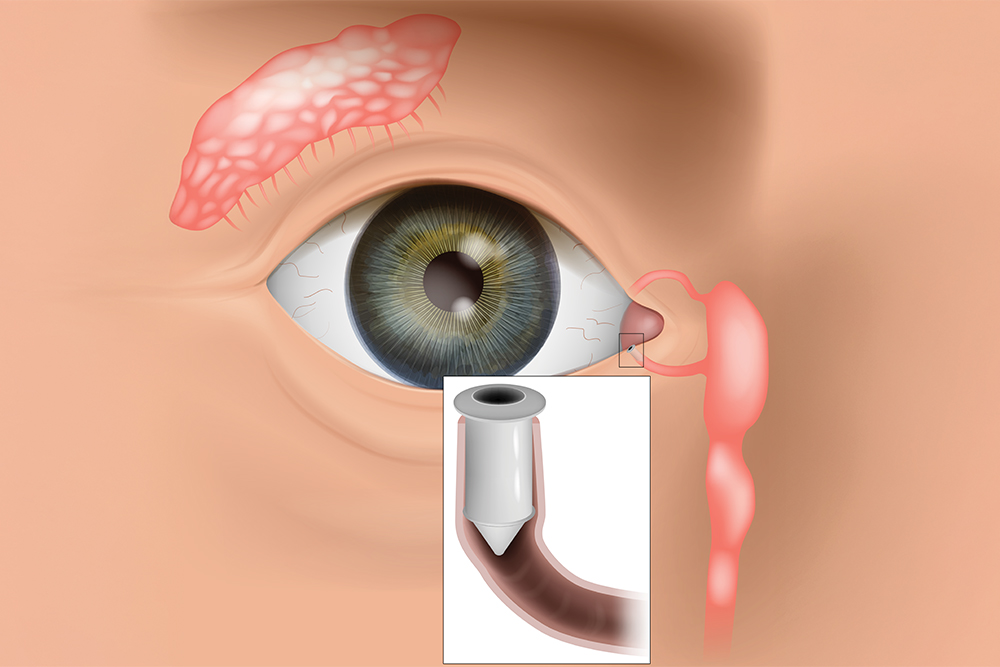
One innovative option designed to provide longer-term relief is LACRIFILL®, a modern approach to punctal occlusion that helps preserve your natural tears and restore balance to the ocular surface.
What Is LACRIFILL® Canalicular Gel?
LACRIFILL® is a soft, clear gel that is placed inside the eye’s natural tear drainage channels, known as the canaliculi. These tiny channels are responsible for carrying tears away from the surface of the eye and into the nasal cavity.
For individuals with dry eye disease, tears often drain away too quickly. When this happens, the eye does not stay properly lubricated, leading to dryness and irritation. LACRIFILL® works by gently filling part of the drainage pathway, slowing tear outflow so that your natural tears remain on the eye longer.
Unlike traditional artificial tears that supplement moisture from the outside, LACRIFILL® helps you retain the tears your body already produces. This treatment is FDA-cleared and has been clinically studied for safety and effectiveness in managing dry eye symptoms.
How Does LACRIFILL® Work?
Every time you blink, a thin layer of tears spreads across the surface of your eye. This tear film keeps your eyes smooth, hydrated, and protected from debris and infection. After serving their purpose, tears drain through small openings in the inner corners of the eyelids (called puncta), passing into the canaliculi and eventually into the nose. In dry eye patients, this drainage process may occur too rapidly, leaving the surface of the eye exposed and under-lubricated.
LACRIFILL® forms a soft gel barrier within the drainage system, reducing tear loss and improving moisture retention. By keeping tears on the eye longer, it supports a healthier tear film and enhances overall comfort.
The gel gradually biodegrades over time and is naturally reabsorbed by the body. Relief typically lasts several months, and the treatment can be repeated when necessary.
Benefits of LACRIFILL®
LACRIFILL® offers several advantages for patients struggling with persistent dry eye symptoms:
FDA-cleared and clinically evaluated for safety and performance
Quick, non-surgical procedure performed in-office
No downtime or recovery period required
Preserves your body’s natural tears rather than relying solely on artificial drops
Provides extended relief that can last for months
Biocompatible material designed to work harmoniously with the body
Customizable treatment approach based on your individual tear drainage system
For patients who are tired of frequent eye drop use, this longer-lasting solution can be life-changing.

Clear Lens Extraction (CLE) is a surgical procedure that involves the removal of the natural lens of the eye and replacing it with an artificial lens implant, also known as an intraocular lens (IOL). CLE is primarily performed to correct refractive errors, such as nearsightedness, farsightedness, and astigmatism.
Understanding CLE and How It Works
Clear Lens Extraction is similar to cataract surgery, where the natural lens is removed and replaced with an IOL. However, in CLE, the lens is clear and not clouded as in the case of cataracts. The procedure begins with the administration of local anesthesia to numb the eye and ensure a painless experience for the patient.
Once the eye is numb, a small incision is made on the cornea, which is the clear front surface of the eye. Through this incision, your eye surgeon accesses the lens and carefully removes it. The artificial IOL is then inserted into the empty lens capsule. The IOL is specifically chosen to correct the patient's refractive error, providing them with improved vision.
After the IOL is implanted, the incision is closed with tiny sutures or self-sealing techniques. Following the surgery, patients are usually prescribed antibiotic and anti-inflammatory eye drops to prevent infection and reduce inflammation.
Factors to Consider Before Undergoing Clear Lens Extraction
Before deciding to undergo Clear Lens Extraction, there are several factors that need to be considered. Firstly, it is crucial to have a thorough eye examination to determine if you are a suitable candidate for the procedure. Your optometrist will evaluate your overall eye health and discuss your expectations and goals for vision correction.
Age is another important factor to consider. CLE is typically recommended for individuals over the age of 40 who have developed presbyopia, a condition that affects near vision. It is also important to have stable vision, as any changes in prescription can affect the accuracy of the IOL power calculation.
Additionally, it is essential to understand the potential risks and complications associated with the procedure. While CLE is generally safe, there is a small risk of infection, bleeding, and retinal detachment. Your optometrist will discuss these risks with you and address any concerns you may have.

In the realm of eye care, surgical co-management has emerged as a collaborative approach that aims to provide patients with comprehensive and seamless treatment. This concept involves the joint efforts of optometrists and ophthalmologists, each bringing their unique expertise to the table. By working together, these eye care professionals strive to enhance patient outcomes and deliver exceptional care.
The Role of Optometrist and Ophthalmologist in Surgical Co-Management
Surgical co-management is built upon the unique skill sets and areas of expertise of optometrists and ophthalmologists. By understanding their respective roles, you can appreciate the synergy that this collaborative approach fosters.
Optometrists are primary eye care professionals who specialize in the examination, diagnosis, and non-surgical treatment of vision disorders. Their responsibilities in surgical co-management include:
Performing comprehensive eye examinations and evaluations
Monitoring and managing pre-existing eye conditions
Providing pre-operative and post-operative care
Educating patients on surgical procedures and aftercare
Collaborating with ophthalmologists to ensure continuity of care
Ophthalmologists are medical doctors who specialize in the diagnosis, treatment, and surgical management of eye diseases and disorders. Their role in surgical co-management encompasses:
Evaluating patients' candidacy for surgical interventions
Performing complex surgical procedures
Providing specialized medical and surgical care
Collaborating with optometrists to ensure seamless patient care
Monitoring and managing post-operative complications
By combining the expertise of optometrists and ophthalmologists, surgical co-management ensures that patients receive comprehensive and coordinated care throughout their treatment journey.
How Surgical Co-Management Works
Surgical co-management is a well-orchestrated process that involves several key steps. Understanding how it works can help you navigate this collaborative approach with confidence.
Initial Evaluation: The process typically begins with an optometrist conducting a comprehensive eye examination. During this evaluation, the optometrist assesses the patient's visual needs, identifies any potential issues, and determines if a surgical intervention is necessary.
Referral and Consultation: If surgery is recommended, the optometrist refers the patient to an ophthalmologist for further evaluation and consultation. This step ensures that the patient receives specialized medical advice and a thorough assessment of their suitability for the proposed surgical procedure.
Pre-operative Care: The optometrist plays a crucial role in providing pre-operative care, which may include managing any existing eye conditions, ensuring the patient understands the surgical process, and addressing any concerns or questions they may have.
Surgical Procedure: The ophthalmologist performs the necessary surgical intervention, leveraging their specialized training and expertise in surgical techniques.
Post-operative Care: After the surgery, the patient's care transitions back to the optometrist, who closely monitors the recovery process and provides post-operative care and management. This may involve follow-up appointments, monitoring for any complications, and ensuring the patient adheres to the prescribed treatment plan.
Ongoing Collaboration: Throughout the entire process, the optometrist and ophthalmologist maintain open communication and collaborate closely. This ensures that the patient's care is seamless, and any concerns or issues are promptly addressed by the appropriate healthcare professional.

Dry eye is a common ocular condition that occurs when the eyes do not produce enough tears or when the tears evaporate too quickly. This can lead to discomfort, redness, blurred vision, and even damage to the surface of the eyes. Understanding the causes and symptoms of dry eye is crucial provide early detection and effective treatment.
The Importance of Dry Eye Advanced Diagnostic Testing
Early detection of dry eye is crucial for preventing further progression of the condition and improving patient outcomes. Without proper diagnosis and treatment, dry eye can cause significant discomfort and affect daily activities. Additionally, chronic dry eye can lead to corneal ulcers, infections, and even vision loss. By accurately identifying and addressing dry eye in its early stages, optometrists can provide timely interventions and prevent complications.
TearLab
One of the advanced diagnostic tools available for dry eye is TearLab. TearLab is a non-invasive test that measures the osmolarity of tears, which is an indicator of tear film stability. This test provides valuable information about the quality and quantity of tears, allowing healthcare professionals to accurately diagnose and monitor dry eye. By analyzing the osmolarity of tears, TearLab helps identify the severity of dry eye and guides treatment decisions. It is a quick and painless procedure that can be performed in a clinical setting.
InflammaDry
Inflammation plays a significant role in dry eye, and identifying the presence of inflammation is crucial for effective treatment. InflammaDry is a diagnostic tool that detects elevated levels of matrix metalloproteinase 9 (MMP-9), an inflammatory marker, in tears. By measuring MMP-9, InflammaDry helps optometrists differentiate between inflammatory and non-inflammatory dry eye. This information is essential for tailoring treatment plans and determining the most appropriate therapies for each patient.

Presbyopia is a natural and inevitable part of aging that affects the eyes' ability to focus on close objects. Unlike other vision conditions, presbyopia isn’t caused by the shape of the eye or structural abnormalities but is instead a result of the eye’s lens losing its flexibility over time.
What is Presbyopia?
Presbyopia is a refractive error, meaning it affects how the eyes bend (or refract) light to focus it on the retina. Unlike nearsightedness (myopia) or farsightedness (hyperopia), which are caused by the shape of the eyeball or cornea, presbyopia is due to the aging of the eye's lens. Over time, the lens becomes less flexible, making it difficult to focus on nearby objects. Presbyopia isn’t a disease or an abnormality—it’s simply a natural consequence of the aging process.
What Causes Presbyopia?
The lens inside the eye changes shape to focus light on the retina, allowing us to see clearly at varying distances. When viewing nearby objects, the lens thickens and curves to increase its refractive power.
As we age, however:
Lens Stiffening: The lens loses elasticity, making it harder to bend and adjust for near vision.
Weakened Ciliary Muscles: The muscles that help change the shape of the lens may lose their strength over time, further reducing the eye’s ability to focus on close objects.
Structural Changes: The lens also grows thicker and less transparent with age, which contributes to reduced focusing ability.
These changes are gradual, typically starting in a person’s 30s and becoming noticeable by their early to mid-40s.

As technology continues to advance, so does the field of the optometric industry. The development of innovative tools and techniques has allowed for more accurate and comprehensive examinations. One such technology is Optos, a revolutionary system that utilizes ultra-widefield retinal imaging technology to provide optometrists with a detailed view of the entire retina.
How Does Optos Work?
Optos technology is based on the principle of ultra-widefield retinal imaging, which allows for a wider and more detailed view of the retina compared to traditional imaging techniques. The Optos system consists of a specialized camera that captures high-resolution images of the retina using scanning laser ophthalmoscopy (SLO) and optical coherence tomography (OCT) technologies. SLO provides a wide-field view of the retina, while OCT allows for cross-sectional imaging, providing valuable insights into the various layers of the retina.
The Optos camera is designed to capture images through a process called optomap, which captures up to 200 degrees of the retina in a single image. This wide-field view provides optometrists with a comprehensive picture of the retina, enabling them to detect abnormalities that may not be visible with traditional imaging techniques. The optomap image can be instantly viewed and analyzed by your eye doctor, allowing for a more efficient and accurate diagnosis.
Common Eye Conditions Detected by Optos
Optos technology has the capability to detect a wide range of eye conditions, including but not limited to, diabetic retinopathy, macular degeneration, glaucoma, and retinal tears or detachments. Diabetic retinopathy is a condition that affects individuals with diabetes, causing damage to the blood vessels in the retina. Optos can capture detailed images of the retina, enabling optometrists to detect any signs of diabetic retinopathy and initiate appropriate treatment.
Macular degeneration is another common eye condition that can be detected using Optos. This condition affects the macula, the central part of the retina responsible for sharp, central vision. Optos allows for a comprehensive view of the macula, identifying any changes or abnormalities that may indicate the presence of macular degeneration.
Glaucoma, a condition characterized by damage to the optic nerve, can also be detected using Optos. The wide-field view provided by Optos allows for a thorough examination of the optic nerve and the surrounding structures, facilitating early detection and intervention.
Finally, Optos technology is particularly effective in detecting retinal tears or detachments. These conditions can lead to sudden vision loss and require immediate medical attention. Optos allows for a comprehensive view of the retina, identifying any signs of retinal tears or detachments and initiate prompt treatment.

Our eyes are extremely delicate, yet they can be subjected to harsh conditions and other environmental factors that affect their health. One of the problems that can affect our eyes is an accumulation of dirt, debris and bacteria on the eyelids. This can cause a range of issues, including stopping tear film from reaching the eyes and being properly dispersed over their surface – which is necessary to keep them healthy and comfortable. Fortunately, a new solution called Blephex® can help.
What is Blephex®?
Blephex® is a handheld electro-mechanical device that is applied to the margins of the eyelids with the purpose of cleaning them and improving the effectiveness with which tear film flows onto the surface of the eyes.
Blephex® has a disposable, surgical-grade sponge tip which rapidly oscillates to create a cleaning action. Before the sponge tip is placed onto the eyes, it is soaked in a gentle exfoliating solution. This solution provides soft abrasion to help remove dead skin cells and debris that could be irritating the eyes and interrupting tear film progression. The Blephex® device is manually applied to the eyes and moved gently across the eyelids, with the entire, painless process taking approximately 6 to 8 minutes per eye. A different sponge is used on each eye, ensuring that no bacteria is passed between them. After the procedure, patients are given instructions on how to maintain the cleanliness of their eyelids with daily/nightly eyelid hygiene at home.
Most patients experience a significant improvement in tear film production and dispersal, and a reduction in unpleasant symptoms that they may have been experiencing within 48 hours of their treatment. While a single treatment is normally enough to produce excellent results, many patients are advised to have Blephex® every 4-6 months.

Dry eye is a common condition that occurs when your eyes do not produce enough tears or when the tears evaporate too quickly. This can result in discomfort, irritation, and a gritty sensation in the eyes. Understanding the causes and symptoms of dry eye is crucial for finding effective treatment options. Tyrvaya offers a breakthrough solution for dry eye relief.
What Causes Dry Eye?
There are several factors that can contribute to the development of dry eye. The meibomian glands are responsible for producing the oily component of the tear film, which helps prevent evaporation of tears and maintains a smooth ocular surface. Meibomian gland dysfunction occurs when these glands become blocked, leading to a decrease in the quantity and quality of the meibum. This can result in evaporative dry eye, discomfort, and inflammation of the eyelid margins.
Another common causes is age. As we get older, our tear production tends to decrease, making us more prone to dryness. Hormonal changes, such as those that occur during menopause, can also affect tear production and lead to dry eye.
Environmental factors can play a role as well. Dry or windy climates, air conditioning, and excessive screen time can all contribute to dry eye. Additionally, certain medications, such as antihistamines and antidepressants, can cause dryness as a side effect.
Other underlying health conditions, such as autoimmune diseases like Sjogren's syndrome or rheumatoid arthritis, can also contribute to dry eye. In these cases, the body's immune system mistakenly attacks the tear glands, leading to reduced tear production.

Optomap is an innovative new technology that gives eye doctors the ability to perform ultra-wide retinal imaging that is far superior to what can currently be achieved using conventional retinal imaging options. In contrast to conventional retinal imaging, Optomap captures at least 50% more of the retina in a single capture, and with Optomap’s multi-capture function, up to 97% of the retina can be viewed. This gives eye care professionals greater opportunity to monitor the health and condition of patient vision.
Why is Optomap important?
Optomap is another great preventative eyecare technology tool. By allowing your eye doctor to have a comprehensive view of your retina, they will be able to detect any developing eye diseases early on, before they have a detrimental impact on your vision and day to day life. Not only can Optomap detect eye conditions such as retinal holes, retinal detachment, macular degeneration and diabetic retinopathy, but it can also be used to identify some general health conditions such as cardiovascular disease, stroke and cancer.
- Monday 8:00am - 5:00pm
- Tuesday 8:00am - 5:00pm
- Wednesday 8:00am - 5:00pm
- Thursday 8:00am - 5:00pm
- Friday 8:00am - 5:00pm
- Saturday 8:00am - 12:00pm
- Sunday Closed
© 2026 Charles Bittel III, O.D., Inc.. All rights Reserved. Accessibility Statement - Privacy Policy - Sitemap
Powered by: